What Is an MCL Injury?
An MCL (medial collateral ligament) tear is a common injury we see in our clinics, especially among those that participate in sports such as football, rugby, or skiing (essentially, any sport that might see sudden changes in direction). It is the most commonly injured of the knee ligaments. The injury involves damage to the medial collateral ligament, situated on the inner side of the knee. The severity of an MCL tear can range from a partial tear (where only some fibres of the ligament are damaged) to complete (where the ligament is entirely split into two pieces).
MCL tears generally occur due to a forceful blow to the knee, or through sudden movements that place excessive strain on the ligament. While surgery may be necessary for severe cases, most MCL tears are successfully treated with non-surgical methods. In this blog, we’ll explore what MCL tears are, their symptoms and causes, and the treatment options available to manage and recover from this injury.
What Is the Medial Collateral Ligament (MCL)?

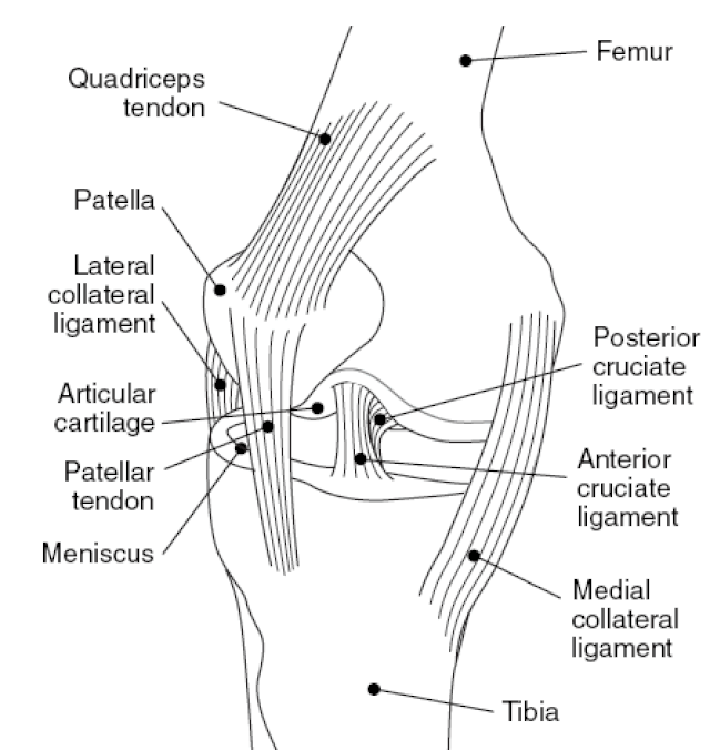
National Institute of Arthritis and Musculoskeletal and Skin Diseases (NIAMS), Public domain, via Wikimedia Commons
The medial collateral ligament, commonly referred to as the MCL, is a vital structure in your knee joint. Located on the inner side of your knee, the MCL spans approximately eight to 10 centimetres in length, connecting the thigh bone (femur) to the shin bone (tibia). It provides strength and stability to the knee and helps maintain alignment and movement during physical activities.
The MCL is one of the four primary ligaments that support the knee. Alongside the MCL, the other three main ligaments include:
- The anterior cruciate ligament (ACL), which helps with rotational stability.
- The lateral collateral ligament (LCL), which stabilises the outer side of the knee.
- The posterior cruciate ligament (PCL), which controls the backward movement of the shin bone.
The term “medial” just means that the MCL is situated towards the middle or centre of the body, while “collateral” signifies its position on one side of the knee joint. Thus, the medial collateral ligament simply means the ligament that is located on the inner side of both knees, closer to the centre line of the body.
Symptoms
The symptoms of a medial collateral ligament (MCL) varies depending on the severity of the injury. Common symptoms include:
- Popping sound – Often, the initial injury is accompanied by a distinct popping sound.
- Pain – You may experience sharp pain in the knee at the time of injury.
- Tenderness – The inner side of your knee may feel tender to the touch.
- Swelling and stiffness – Swelling is common and can contribute to a stiff feeling in the knee.
- Instability – There might be a sensation that the knee is about to “give out” under weight-bearing pressure.
- Locking or Catching – The knee joint may lock or catch during movement.
Immediate medical attention is advisable if you experience these symptoms, even if walking is still possible, to prevent further damage and begin appropriate treatment.
The Types of Medial Collateral Ligament Injury
MCL injuries are classified into three grades, reflecting the severity of the tear:
Grade 1
- Description – This is considered a mild tear where less than 10% of the fibres in the MCL are torn.
- Symptoms – Individuals with a Grade 1 tear typically experience tenderness along with mild pain around the area of the injury. However, the knee remains stable, and the overall integrity of the joint is not compromised. Walking is typically still possible, though it may be accompanied by pain.
Grade 2
- Description – This is a moderate tear that usually involves a partial tearing of the MCL, often affecting the superficial part of the ligament.
- Symptoms – The knee may feel loose when manipulated by hand. There is usually intense pain and tenderness located on the inner side of the knee. The stability of the knee is compromised but not completely lost. Walking can be difficult due to decreased stability in the knee.
Grade 3
- Description – This is a severe injury where the MCL is completely torn, affecting both the superficial and deeper parts of the ligament.
- Symptoms – A Grade 3 tear leads to significant instability in the knee, with the joint feeling very loose. Intense pain and tenderness are common, and the injury is frequently associated with damage to other knee structures, such as the anterior cruciate ligament (ACL). It becomes very challenging to walk due to significant instability and intense pain. Walking might be avoided entirely to reduce discomfort.
Each grade requires a different approach to ensure optimal recovery and to prevent further damage to the knee joint.
Causes
MCL tears are generally caused by actions that place excessive stress on the knee:
- Cutting movements – Planting the foot and making a sharp turn, common in sports, often leads to tears.
- Direct impact – A blow to the outer side of the knee, such as from a tackle in football, can cause the ligament to stretch or tear.
- Awkward landings – Landing improperly from a jump can stress the knee.
- Hyperextension – Overextending the knee, a frequent occurrence in skiing, can damage the MCL.
- Chronic stress – Continuous pressure on the knee from activities like squatting or repeated stress in sports can wear down the ligament over time.
MCL injuries are particularly common among athletes engaged in sports with sudden movements and physical contact. Understanding these causes and symptoms helps in the prevention and early detection of MCL injuries, enabling timely and effective treatment strategies.
Diagnosis
When you visit your doctor with a knee injury, they will begin by gathering detailed information about how the injury occurred and your subsequent symptoms and mobility challenges. Understanding the circumstances of your injury and your experiences since then helps in forming an initial assessment.
Physical Examination
Your doctor will conduct a thorough physical examination to assess the severity and extent of the MCL injury. This examination may include:
- Palpation – Applying pressure on the inside of your knee to check for pain and stability. This helps determine how loose or stable the joint is.
- Movement tests – Pressing on the outside of your knee while moving your leg in various positions—both bent and straight—to evaluate the extent of the injury.
Imaging Tests
To gain a clearer picture of the injury, your doctor may recommend several imaging tests:
- MRI (Magnetic Resonance Imaging) – This is particularly useful as it provides detailed images of both hard and soft tissues, allowing your doctor to see the extent of the MCL damage and any other associated injuries within the knee.
- X-ray – While an X-ray does not show soft tissues like ligaments, it is valuable for ruling out any fractures that might accompany the ligament injury.
- Stress X-ray – This specific type of X-ray assesses the stability of the MCL. During the test, while you relax your leg, a technician will apply gentle pressure to the side of the knee associated with the MCL. If the images show an unusually wide gap between the thigh and shin bones, it suggests that the joint is loose, indicating a potential MCL tear.
- Ultrasound – Using sound waves, an ultrasound can generate real-time images of the internal structures of your knee. This test is useful for assessing the severity of the MCL tear and checking for other damages to the knee.
These diagnostic steps are crucial for determining the most effective treatment plan. By accurately assessing the condition of the MCL and other structures in the knee, your doctor can tailor a treatment strategy that optimises healing and facilitates a return to normal function. They should let you know what grade of tear you’ve suffered.
Treatment
The first step to treatment is getting an official diagnosis. If you think you’ve injured your MCL, be sure to see a medical professional. If the pain is severe, and especially if it’s challenging to walk, go to the emergency room.
Initial steps
The immediate treatment goal is to manage pain and swelling effectively:
- RICE protocol – Rest, Ice, Compression, and Elevation should be your first courses of action.
- HARM avoidance – Heat, Alcohol, Running (or any strenuous activity), and Massage on the injured area should be avoided as they can exacerbate swelling and pain.
Medication
- Over-the-counter pain relief – Medications like paracetamol or ibuprofen are commonly used to alleviate pain and reduce inflammation.
- Prescription painkillers – For more severe pain, your doctor might prescribe stronger painkillers.
- NSAIDs – Non-steroidal anti-inflammatory drugs help to reduce inflammation along with pain relief. Always consult healthcare providers for appropriate dosages and potential side effects.
Physiotherapy
Physiotherapy is a fantastic choice when looking to treat an MCL injury. A physiotherapist will:
- Assess – Conduct a thorough assessment of your knee’s condition.
- Plan – Develop a personalised rehabilitation program that includes exercises aimed at restoring range of motion, strength, and stability to the knee.
- Educate – Provide guidance on how to perform exercises correctly and safely to aid recovery and prevent future injuries. Physiotherapy is often all that is needed for recovery, especially in cases of MCL sprains where the ligament is not completely torn.
If you’re in Sydney and need an experienced physiotherapist for your MCL injury, contact us today. Otherwise, why not book one of our online physiotherapy appointments?
Knee Brace
Wearing a knee brace can stabilise the knee and prevent movements that could aggravate the injury. This is often used in conjunction with physiotherapy.
Surgery
While most MCL injuries heal with non-surgical treatment, surgery might be necessary if:
- Multiple injuries – More than one ligament or significant structural damage is involved.
- Persistent instability – If the knee remains unstable after extensive physiotherapy. Surgical intervention usually involves repairing or reconstructing the damaged ligament to restore the knee’s stability.
Consultation with Specialists
You may be advised to see an orthopaedic surgeon or a sports medicine professional depending on the complexity of your injury. These specialists can offer further advice and treatment options, ensuring you receive the best care for your specific needs.
Contact Benchmark Physio for Your Physiotherapy Needs
While an MCL injury may be serious, it doesn’t have to mean the end of your sporting career or hobby. Contact us today for a personalised physiotherapy treatment plan and we’ll get you back to doing what you love in no time.


